Structures of the ear and hearing
- Aarti Makan
- Sep 3, 2019
- 4 min read
Updated: Feb 21, 2020
Index
The ear
The outer ear
Pinna
Ear canal
Ear drum
The middle ear
Ossicles
Eustachian tube
The ear’s protective reflex
The inner ear
Cochlea
The acoustic nerve and the brain
Acoustic nerve
The brain
The ear
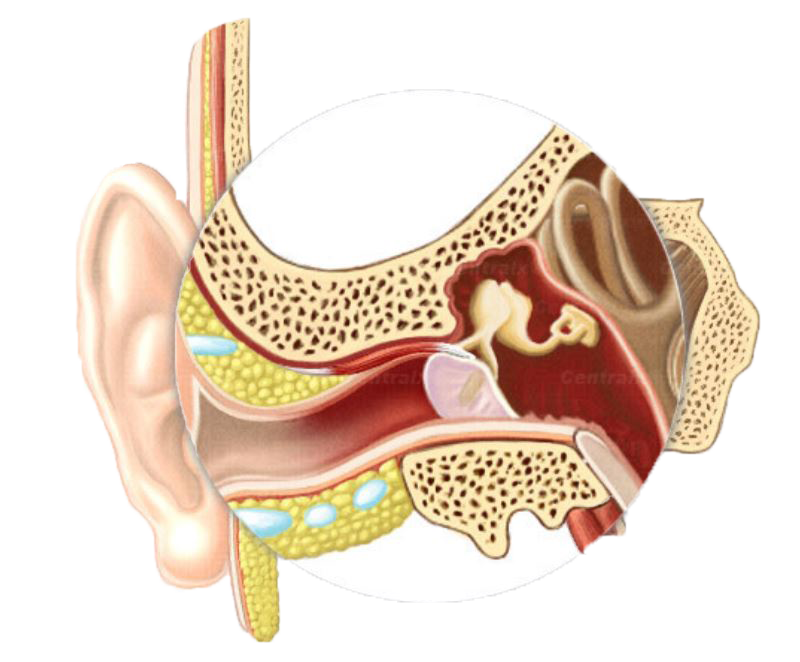
The ear is the part of the body responsible for hearing and balance. Only vertebrates (animals with backbones) have ears. Invertebrate animals, such as jellyfish and insects, lack ears, but have other structures or organs that serve similar functions. The most complex and highly developed ears are those of mammals. The ear does not work in isolation, it transmits sounds via the auditory nerve to the brain. The human ear consists of three sections: the outer, middle, and inner ear. The outer and middle ears function only for hearing, while the inner ear is also responsible for your balance and orientation.

The outer ear
The outer ear is made up of the pinna, the external ear canal and the outer part of the eardrum.


Pinna
The pinna is a cartilaginous part of the ear attached to the side of the head. It acts as a funnel that collects sounds from the environment and transfers it into the ear canal.

Ear Canal
The ear canal is a tube-like passage that measures about 3cm in length. It leads from the pinna to the eardrum. The first part of the ear canal is lined with delicate hairs and small glands that produce wax. Wax prevents dust and dirt from entering the deeper part of the ear. The inner two-thirds of the ear canal is surrounded by the temporal bone of the skull, which also surrounds the middle and inner ear. The temporal bone protects these fragile parts of the ear.

Eardrum
The eardrum separates the outer ear from the middle ear. It is a thin, round, skin-like membrane that is stretched tight like a drum across your ear canal. When a drummer beats a drum, the drum vibrates to make sounds – similarly, sound hits the eardrum and causes it to vibrate. The eardrum amplifies the vibrations of sound so it can travel from the tiny bones of the middle ear, which then sends the vibrations into the inner ear.
The middle ear
The middle ear is a narrow, air-filled chamber about 1.5cm cubed in size. The middle ear has 3 main parts: the inner part of the eardrum, the ossicles and the eustachian tube.


Ossicles
The inner part of the eardrum is attached to the middle ear by the first bone of the ossicular chain, the malleus. The ossicles are the tiniest bones in your body - each about the size of a grain of rice. They each have a unique name based on the shape; the malleus or hammer, the incus or anvil and the stapes or stirrup. These 3 bones are responsible for transferring sound from the eardrum to the inner ear. This is because the last bone, stapes, is attached to the cochlea. These bones also have a function to protect the ear.

Eustachian tube
A narrow passageway called the eustachian tube connects the middle ear to the throat and the back of the nose. The eustachian tube helps keep the eardrum intact by equalising the pressure on either side of the middle and outer ear. For example, if you are in an airplane while taking off, the air pressure becomes lower as the plane climbs higher in altitude. Your ears may feel some discomfort at this point because the air pressure in the middle ear becomes greater than the pressure in the outer ear. When you yawn or swallow, the eustachian tube opens, and some of the air in the middle ear passes into the throat, adjusting the pressure in the middle ear to match the pressure in the outer ear. This equalising of pressure on both sides of the eardrum prevents it from rupturing.
The ear’s protective reflex
An acoustic reflex is the protective mechanism of your ear. When loud noises produce forceful vibrations, two small muscles called the tensor tympani and the stapedius, contract and limit the movement of the ossicles. This is similar to when you instinctively pull your hand away from a hot plate. This action reduces the intensity of the sound that enters the main organ of hearing (cochlea) and thus reduces damage to your ears.


The inner ear
The inner ear contains the main organs of hearing (cochlea) and balance (the vestibule and the three semi-circular canals)


Cochlea
The cochlea is the main organ of hearing. It is a coiled tube about the size of a pea and looks like the shell of a snail.

The cochlea is filled with fluid and within this tube are rows of tiny sensors or hair cells responsible for hearing. These hair cells give us the ability to discriminate between sounds of different pitch or frequency.
The acoustic nerve and the brain

Acoustic nerve
This is the nerve that is responsible for hearing, balance, and head position. Its primary function is to transmit sounds from the inner ear to the brain. It has two parts: a cochlea part (auditory) that transmits sound reception for hearing and a vestibular part (balance) that senses balance and head position.

The brain
It is in the brain that sounds are identified, processed and understood (auditory processing). The part of the brain responsible for auditory processing is called the temporal lobe. Both ears work together to identify sounds from all around us. This gives the brain the full information in order to identify the type of sound and which direction it is coming from.
The temporal lobe is known as the primary auditory cortex (the first part of the brain that receives sounds from the ear). It is the main part of the brain responsible for understanding written language, spoken language and speech discrimination. It also maintains long-term memory.
Learn how the structures of the ear work with our interactive animation.
Watch video about how humans hear:

Comments